Radiologically Inserted Gastrostomies (RIG)
Safe, Image-Guided Feeding Tube Placement
Overview
Radiologically Inserted Gastrostomy (RIG) is a minimally invasive procedure used to place a feeding tube directly into the stomach under image guidance (X-ray or fluoroscopy).It is commonly performed for patients who are unable to eat or swallow safely, ensuring adequate nutrition and hydration without the need for major surgery.
What is a Gastrostomy?
A gastrostomy is a medical procedure in which a tube is inserted through the abdominal wall into the stomach to provide direct nutritional support.RIG is performed by an interventional radiologist using imaging techniques, making it a safer alternative to surgical methods.
Who Needs RIG?
RIG is recommended for patients who:
- Have difficulty swallowing (dysphagia)
- Have neurological conditions (e.g., stroke, Parkinson’s disease)
- Have head and neck cancers
- Require long-term nutritional support
- Are unable to maintain adequate oral intake
Benefits of RIG
- Minimally invasive (no major surgery)
- Safe and precise placement using imaging
- Suitable for high-risk or fragile patients
- Quick procedure (usually under 1 hour)
- Short hospital stay
- Reliable long-term nutritional support
How the Procedure Works
1. Pre-Procedure Evaluation
Blood tests and imaging are done to assess suitability.
2. Preparation
The stomach is inflated using a tube to allow safe placement.
3. Image-Guided Insertion
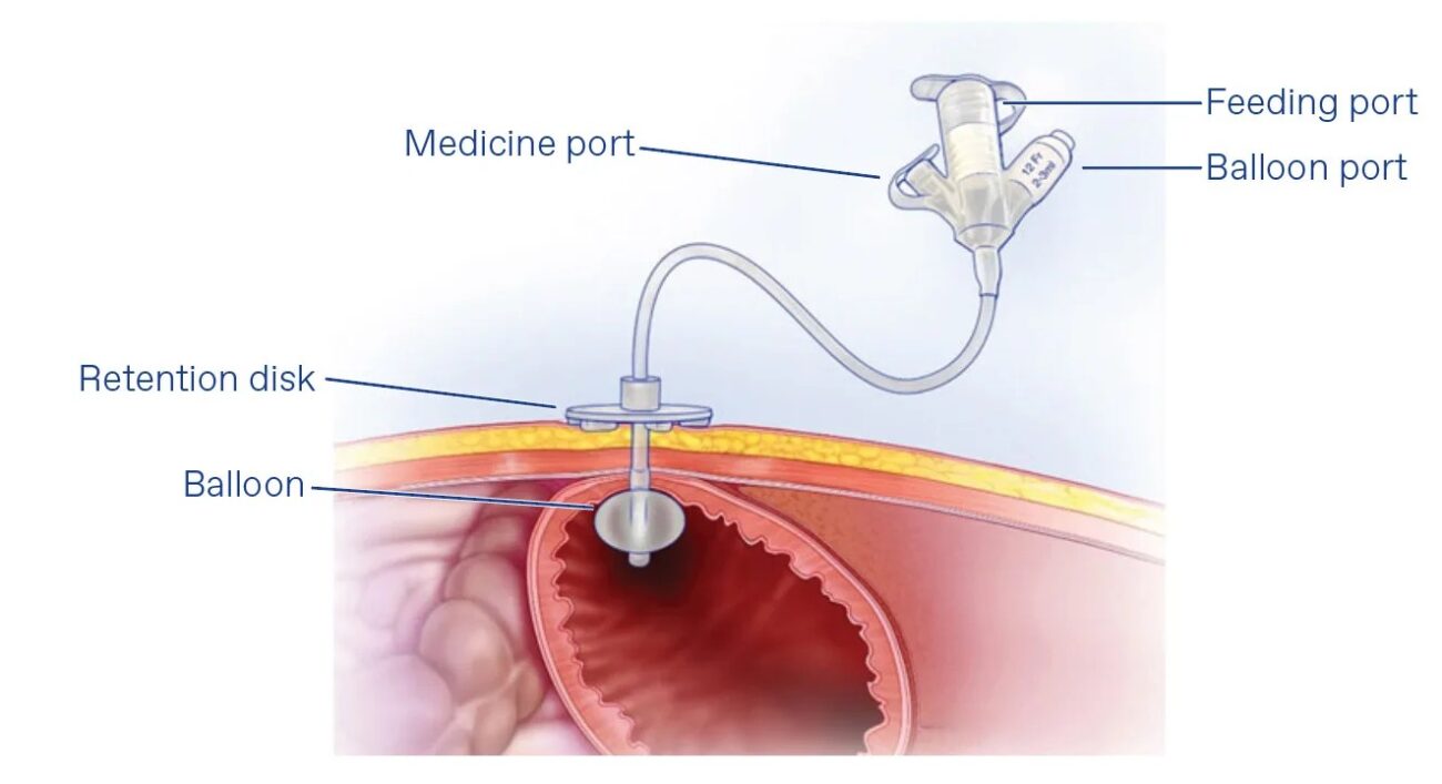
A small incision is made, and the feeding tube is inserted into the stomach using fluoroscopic guidance.
4. Securing the Tube
The tube is secured in place, and a dressing is applied.
Recovery & Aftercare
- Usually requires a short hospital stay
- Feeding can begin within 24 hours (as advised)
- Proper tube care and hygiene are essential
- Caregivers are trained for feeding and maintenance
- Regular follow-up for tube function
Risks & Possible Complications
RIG is generally safe, but possible risks include:
- Mild pain or discomfort at the insertion site
- Infection (rare)
- Tube blockage or displacement
- Leakage around the tube
Your doctor will discuss all risks before the procedure.
Why Choose Us?
- Experienced Interventional Radiology team
- Advanced imaging-guided techniques
- Safe and patient-focused care
- Comprehensive support for patients and caregivers
FREQUENTLY ASKED QUESTIONS
The procedure is done under local anesthesia and sedation, so discomfort is minimal.
Usually 30–60 minutes.
Typically within 12–24 hours after placement, as advised by the doctor.
It can be temporary or long-term depending on the patient’s condition.
Yes, if the patient regains the ability to eat normally.